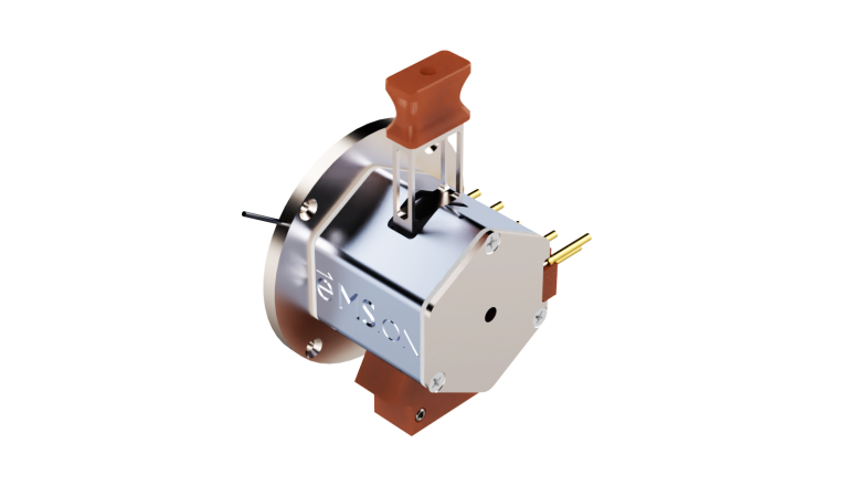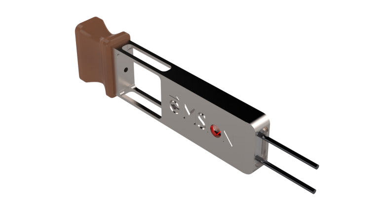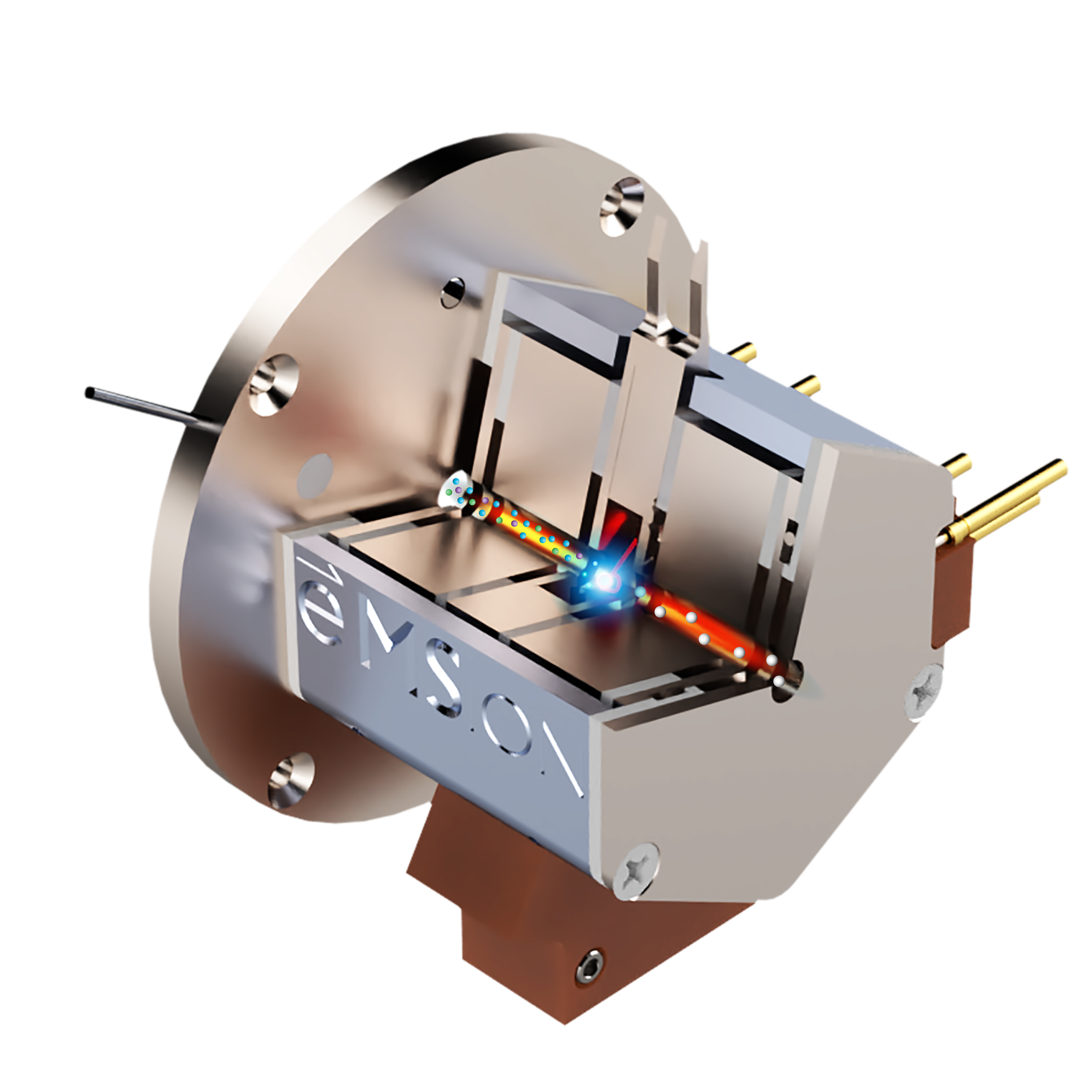
PERMANENT RING MAGNET
FILAMENT
ELECTROSTATIC LENSES
Increase sequence coverage of larger peptides and intact proteins beyond the limits of CID alone.
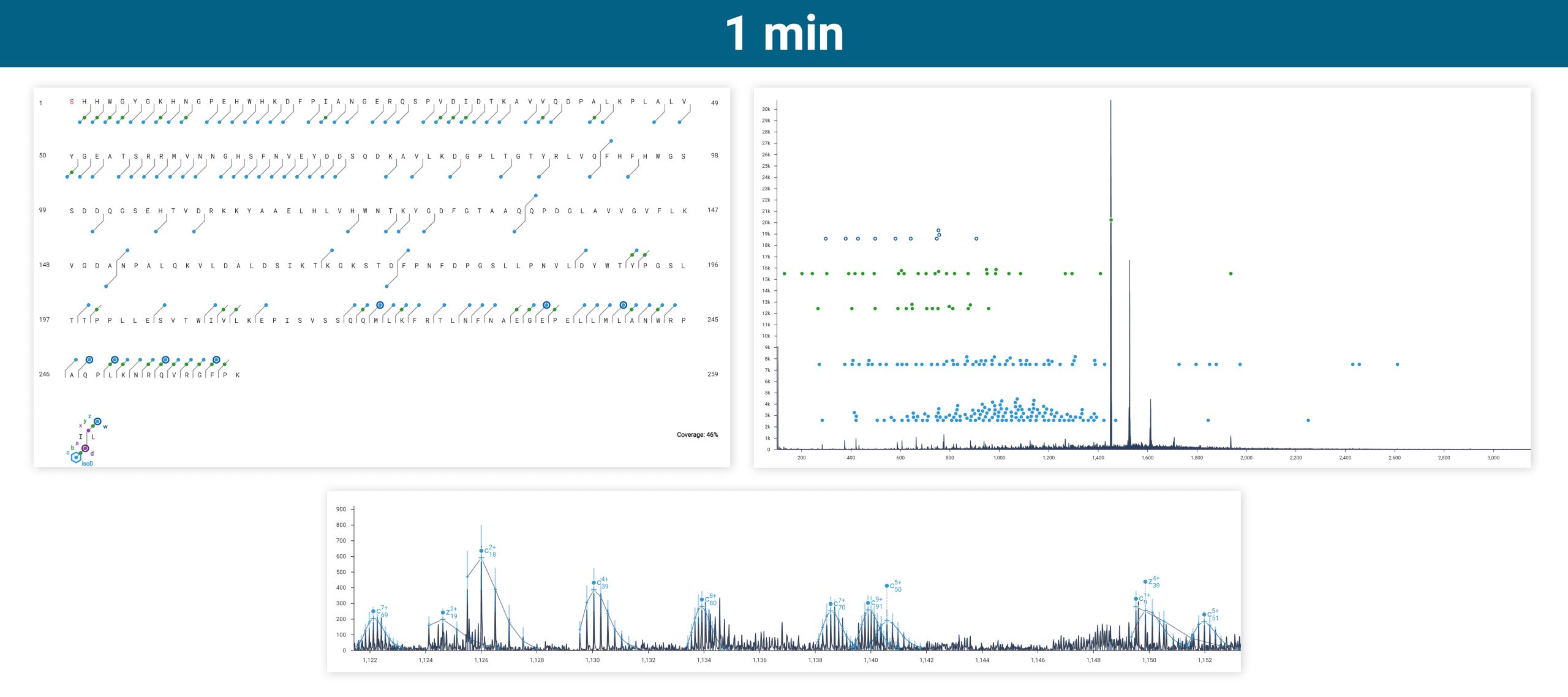
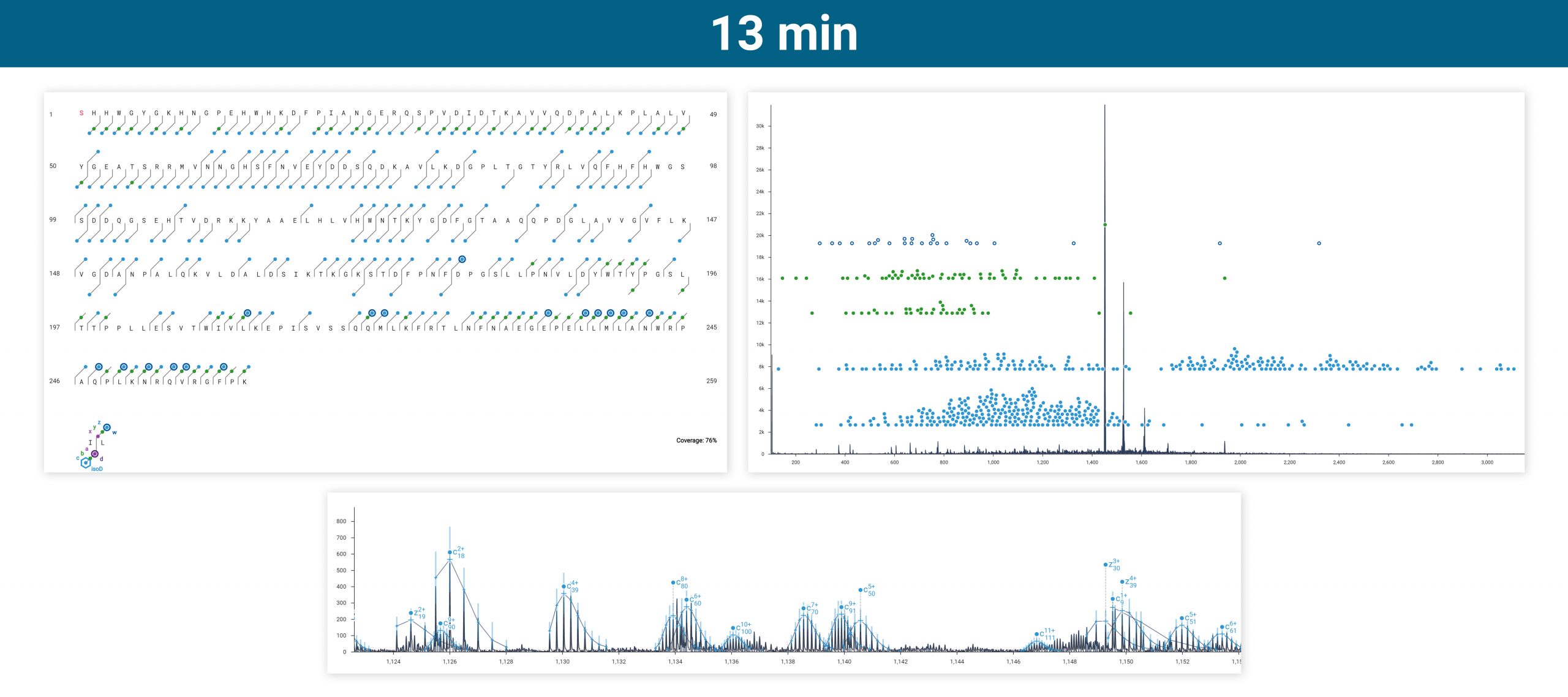
Intact carbonic anhydrase II was run via infusion on an Agilent 6545XT equipped with the ExD option. Top-down ECD was performed on the isolated 20+ charge state, producing amino acid sequence coverage of 28-76% depending on how many acquisitions were averaged.
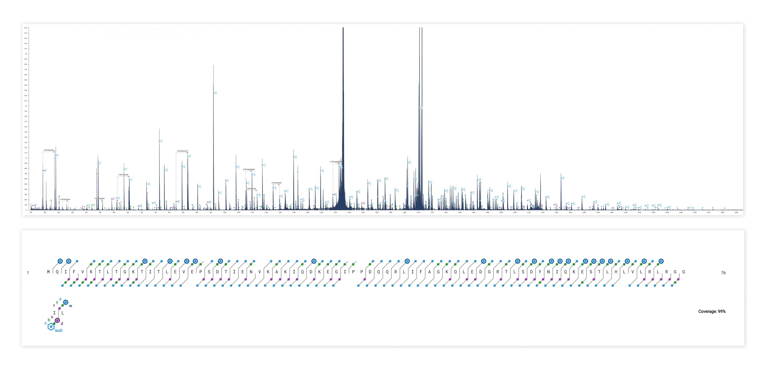
ECD produces sequence-informative fragments for proteins and large peptides while avoiding the non-informative fragmentation that can occur via CID or UVPD. Top-down ECD of 6+ ubiquitin in the 6545XT AdvanceBio LC/Q-TOF yielded a complete sequence of fragment ions, minus the N-terminus of proline residues, which are not dissociated by ECD.
Side chain fragments (w-ions) produced by ECD can be used to distinguish isobaric amino acids.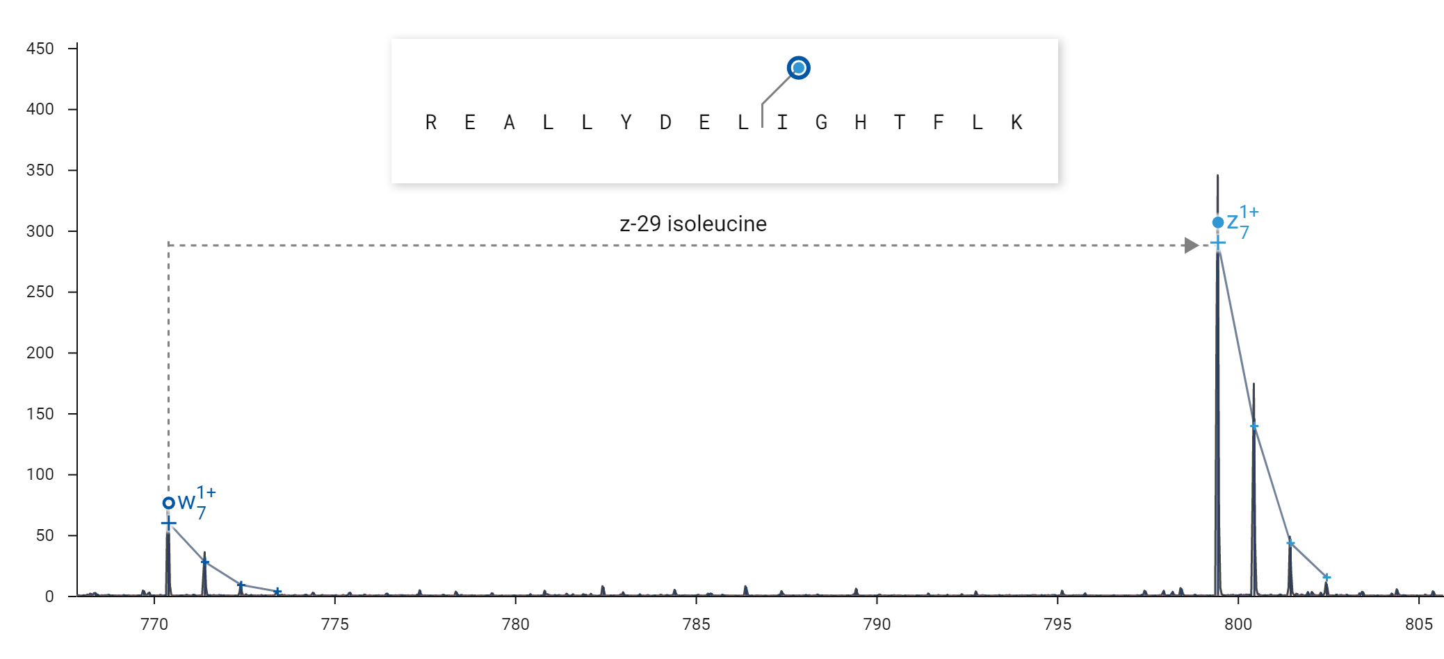
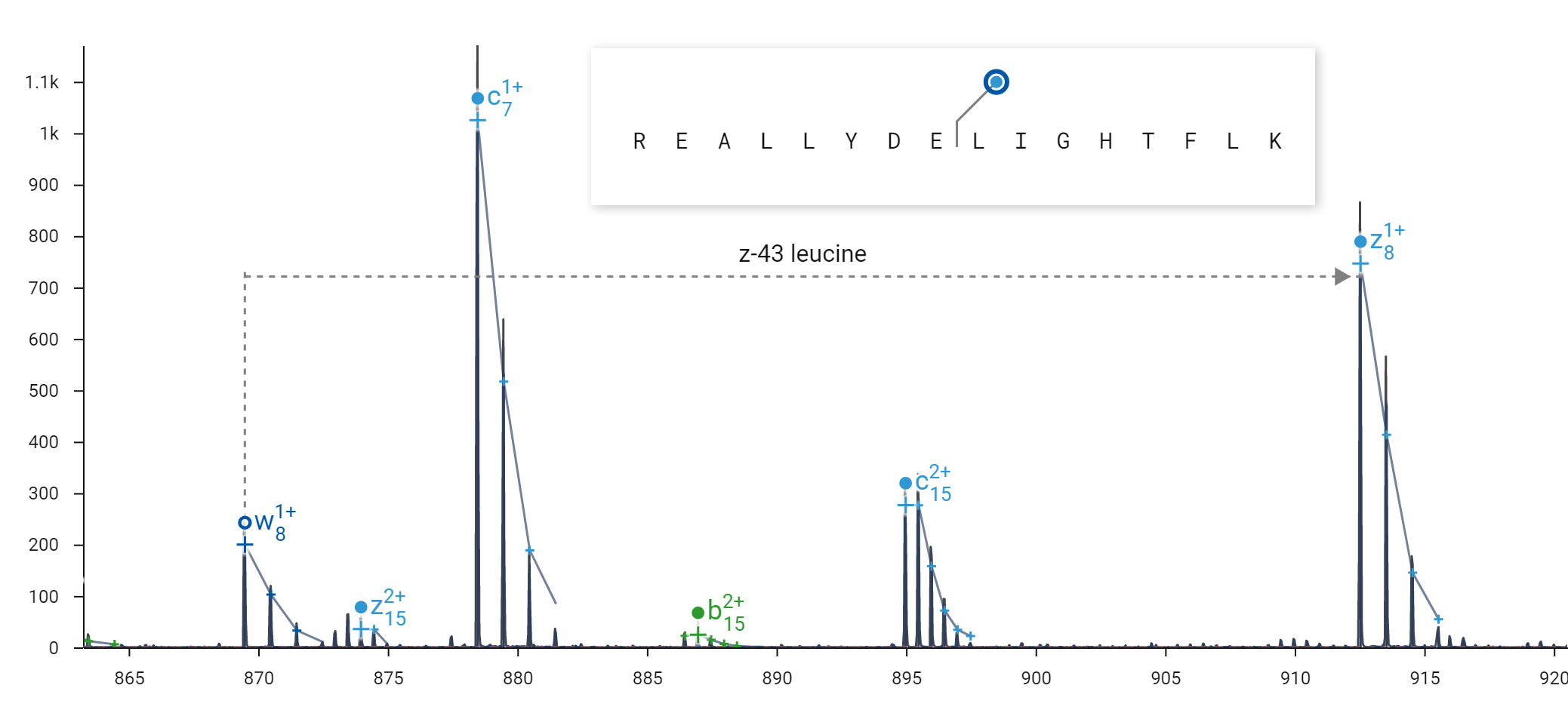
ECD can produce side-chain fragmentation useful for distinguishing leucine from isoleucine residues. ECD was performed on the 2+ peptide REALLYDELIGHTFLK in an Agilent 6545XT Q-TOF. Top: The diagnostic loss of 29 Da from the z7 fragment indicates the presence of isoleucine at residue 10. Bottom: The diagnostic loss of 43 Da from the z8 fragment indicates the presence of leucine at residue 9.
ECD fragmentation products unique to isoaspartate clearly differentiate it from the non-isomerized form. Isoaspartate is implicated in age-related protein inactivation and aggregation, and reduced efficacy of protein therapeutics.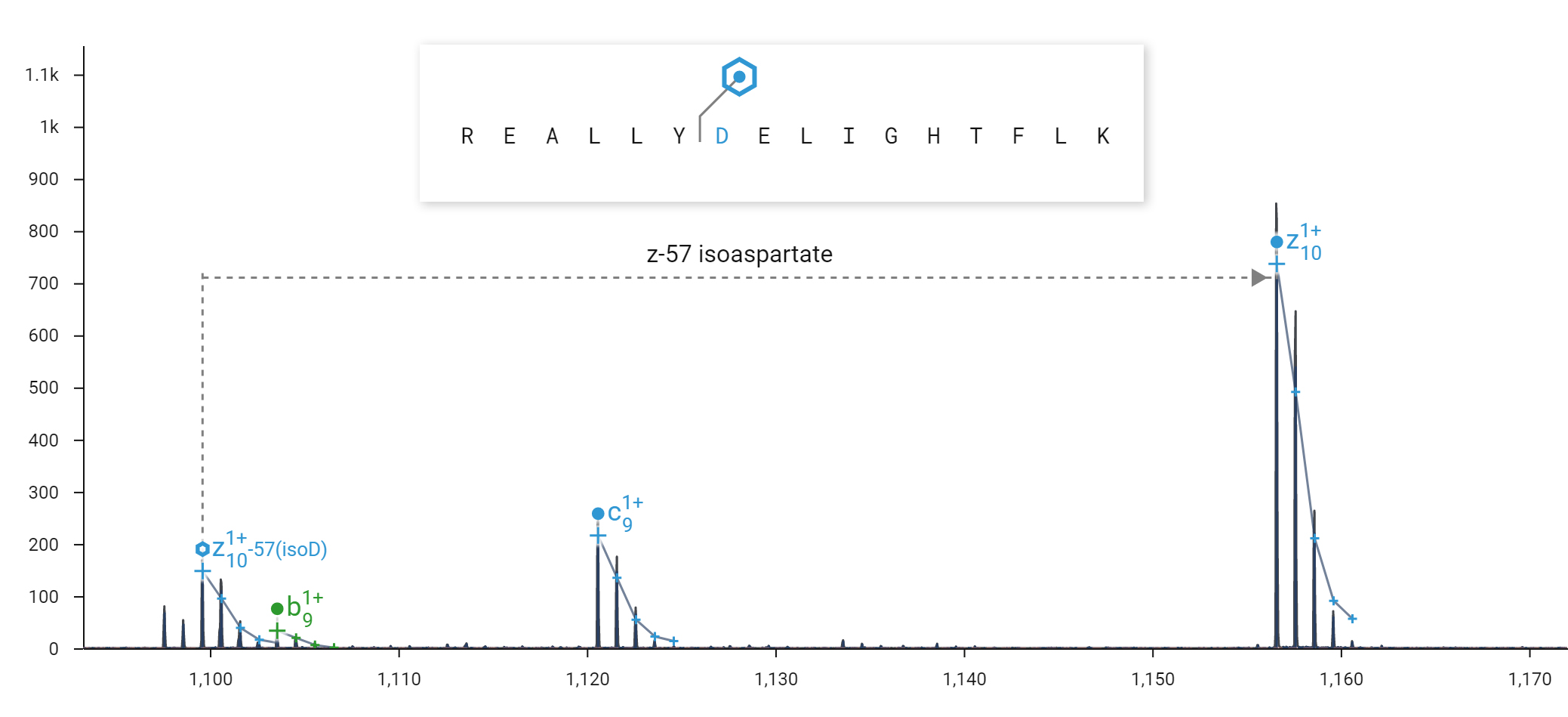
ECD can produce fragment ions diagnostic for isoaspartate. ECD was performed on the 2+ peptide REALLYDELIGHTFLK in an Agilent 6545XT Q-TOF. A 57 Da loss (-C2HO2) from the z10+ ion of the peptide REALLYisoDELIGHTFLK is diagnostic for isoaspartate at position 7. A loss of 45 Da (-CHO2) would be diagnostic for aspartate.
ECD is a “gentle” fragmentation method that preserves labile PTMs – phosphorylation, glycosylation – on fragment ions. With the ExD cell installed, use ECD to map their precise location on peptides and proteins.
Click here to download the Application Note for a demonstration of phosphopeptide analysis using e-MSion’s ExD Cell in an Agilent 6550 iFunnel™ Q-TOF.
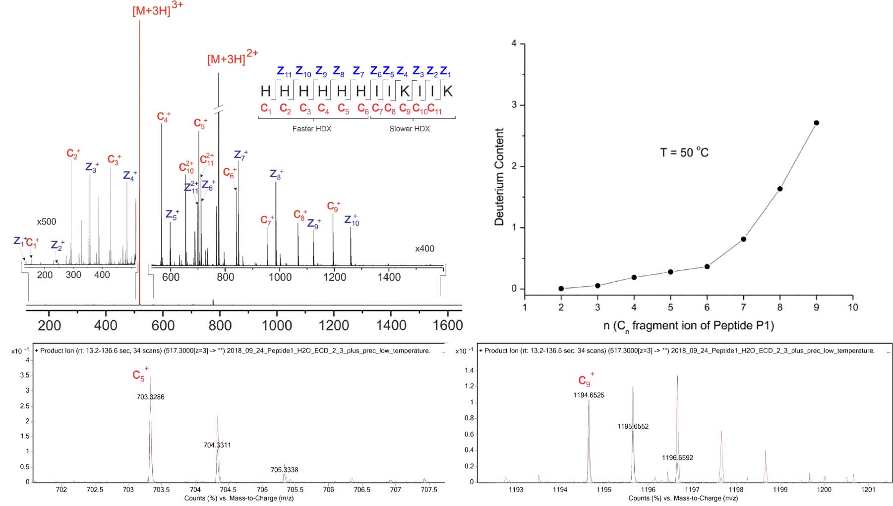
The ExD option enables single-residue-resolution HX-MS with minimal hydrogen scrambling. Top Left: ECD product ion mass spectrum of the [M+3H]3+ precursor of the HX-MS model peptide “P1”, acquired using the ExD Cell in an Agilent 6545 Q-TOF. The His-rich N-terminal half of P1 is engineered to exchange hydrogen more quickly than its C-terminal half. Following back-exchange of deuterated P1, N-terminal c fragments are expected to retain relatively little deuterium, while C-terminal c fragments are expected to retain more deuterium. Any deviations from this pattern would be indicative of hydrogen migration, or “scrambling”, as a result of vibrational excitation. Top Right: After allowing P1 to back-exchange for 5 minutes, deuterium content by residue was similar to expected results published by Rand et al. JACS 130: 1341 (2008): N-terminal residues exchanged hydrogen more quickly than C-terminal residues. Bottom Left: The isotopic distribution of back-exchanged (purple) c5 fragment is similar to protonated (black). Bottom Right: The isotopic distribution of back-exchanged (purple) c9 fragment is shifted right relative to protonated (black), indicating residual deuterium incorporation.
Selective dissociation of disulfide bonds by ECD enables their localization without prior sample reduction.
Figure 1 – Insulin structure and fragments identified in an LC/ECD experiment (average of 5 scans). c– and z-type ECD ions are indicated with blue dots, green dots indicate b– and y-type ions, and purple dots signify a-type ions. Circled dots indicate w-ions. Top: fragments found with Cys7-Cys7 disulfide bridge assumed to be intact. Each chain was calculated separately. The fragments of the B-chain were calculated with the mass of the A-chain as a modification on Cys7, and fragments of the A-chain were calculated with the mass of the B-chain on its Cys7. Bottom: fragments found with the Cys19-Cys20 disulfide bridge intact. The fragments were calculated in the same way as the upper figure, with the mass of the A-chain on Cys19 of the B-chain and the mass of the B-chain on Cys20 of the A-chain.
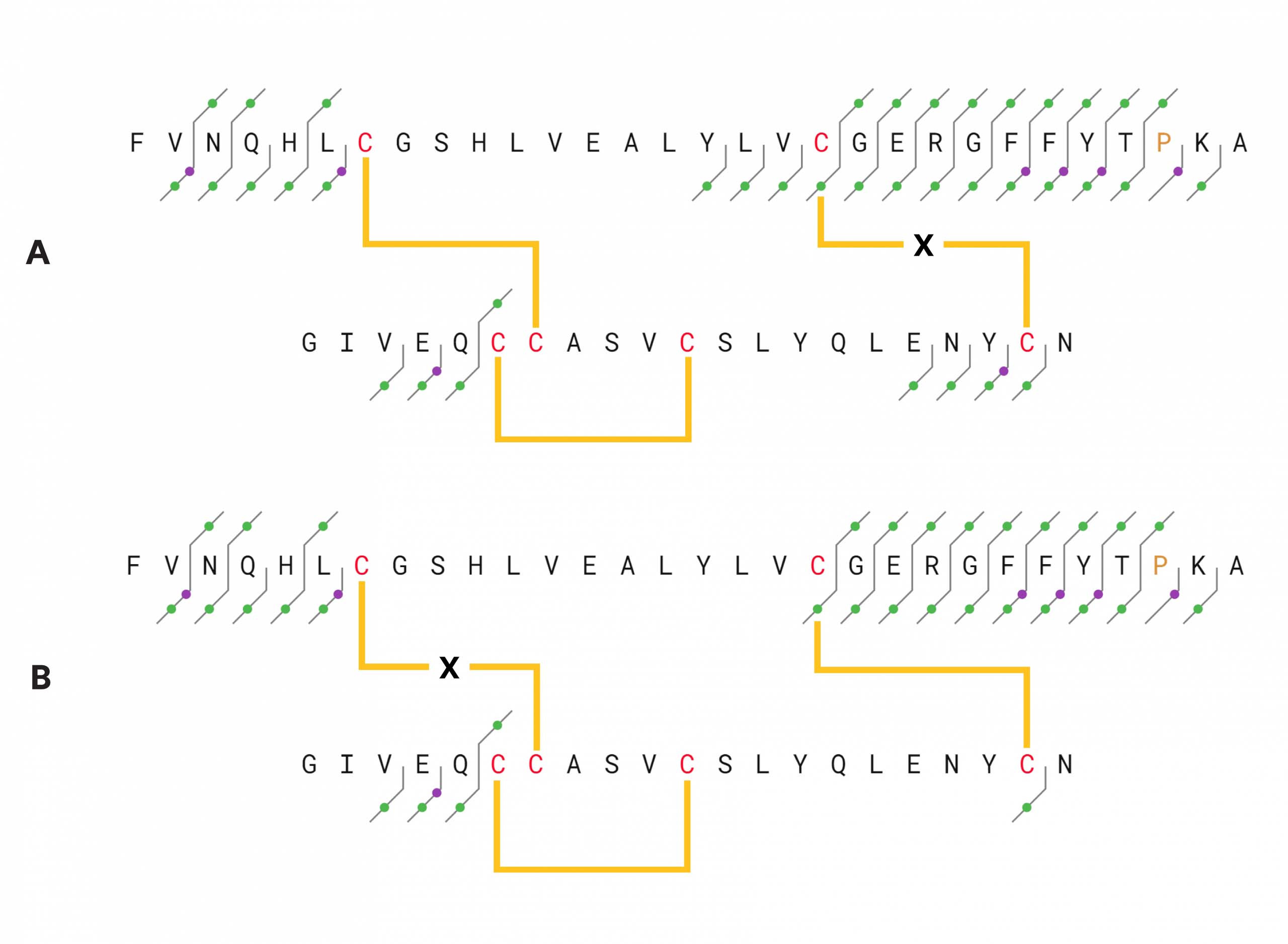 Figure 2 – Insulin structure and fragments identified by CID, 25 V collision energy.
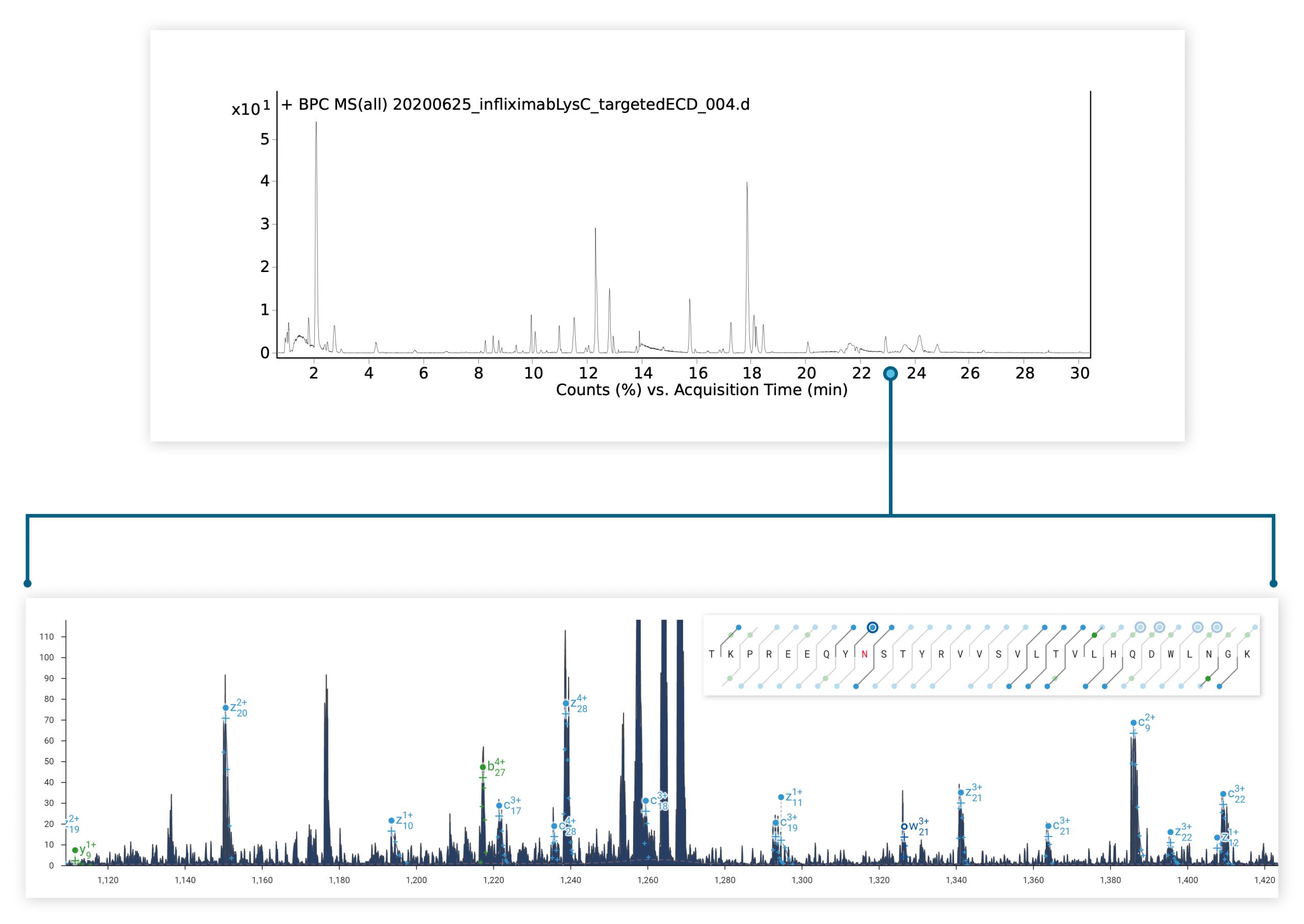
The ExD option is compatible with HPLC timescales and adds valuable information to bottom-up peptide mapping experiments. Here, ECD was used in a targeted manner to fragment a glycopeptide from a Lys C digest of a mAb. The zoomed-in spectrum shows a series of z ions confirming the site & composition of the N glycan without fragmenting the glycan itself.
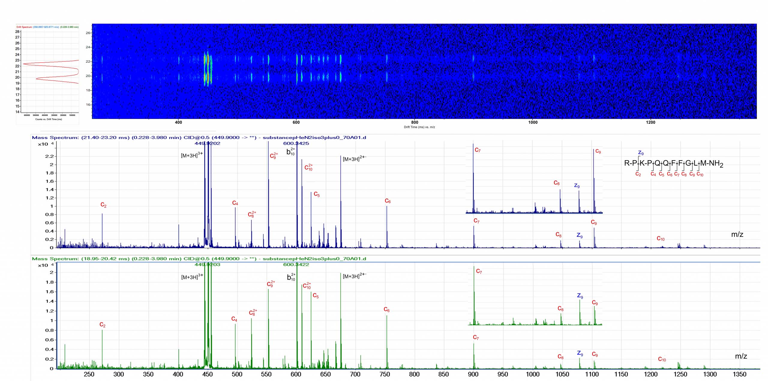 The ExD option is compatible with ion mobility separation in the Agilent 6560 Ion Mobility LC/Q-TOF. Top: Ion Mobility drift spectrum revealing two conformers of [M+3H]3+ Substance P. Bottom: The ExD cell was used to generate ECD product ion mass spectra of the two different Substance [M+3H]3+ conformers. Data courtesy of Dr. Cathy Costello, Boston University.
The ExD option is compatible with capillary electrophoresis timescales. Top: BPC CZE electropherogram of a mixture of five intact proteins. Bottom: ECD sequence coverage maps for two of the proteins in the mix. Data courtesy of Liangliang Sun, Michigan State University.
Install the ExD cell in your current instrument to maximize your investment, or add ExD to an instrument purchase order to capitalize on the increased sensitivity and resolution of newer technology.
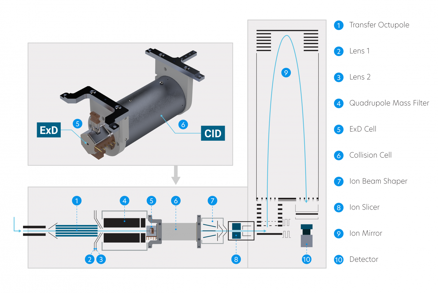
The ExD cell attaches to the entrance of a shortened collision cell, replacing the original collision cell during installation. This way, ExD occurs after isolation by the quadrupole mass filter without affecting collision cell operation.
The ExD Option is available for Agilent 6545XT AdvanceBio and 6560 Ion mobility LC/Q-TOF.
Our ExD filament design features a plug-in cassette with exchangeable filament insert for quick and easy replacement.
ExD is the only electron fragmentation technology fast enough to work after IMS. The ExD cell operates at speeds greater than the fastest separation technique, and has been successfully tested with HPLC, IMS, and CE.

ECD is a “gentle” fragmentation method that preserves labile PTMs – phosphorylation, glycosylation – on fragment ions. With the ExD cell installed, use ECD to map their precise location on peptides and proteins.
Selective dissociation of disulfide bonds by ECD enables their localization without prior sample reduction.
The ExD cell can operate in either ECD or transmission-only mode.
Now, the ExD Software enables automatic switching between selected ExD modes in sync with changes in instrument scan type during data acquisition.